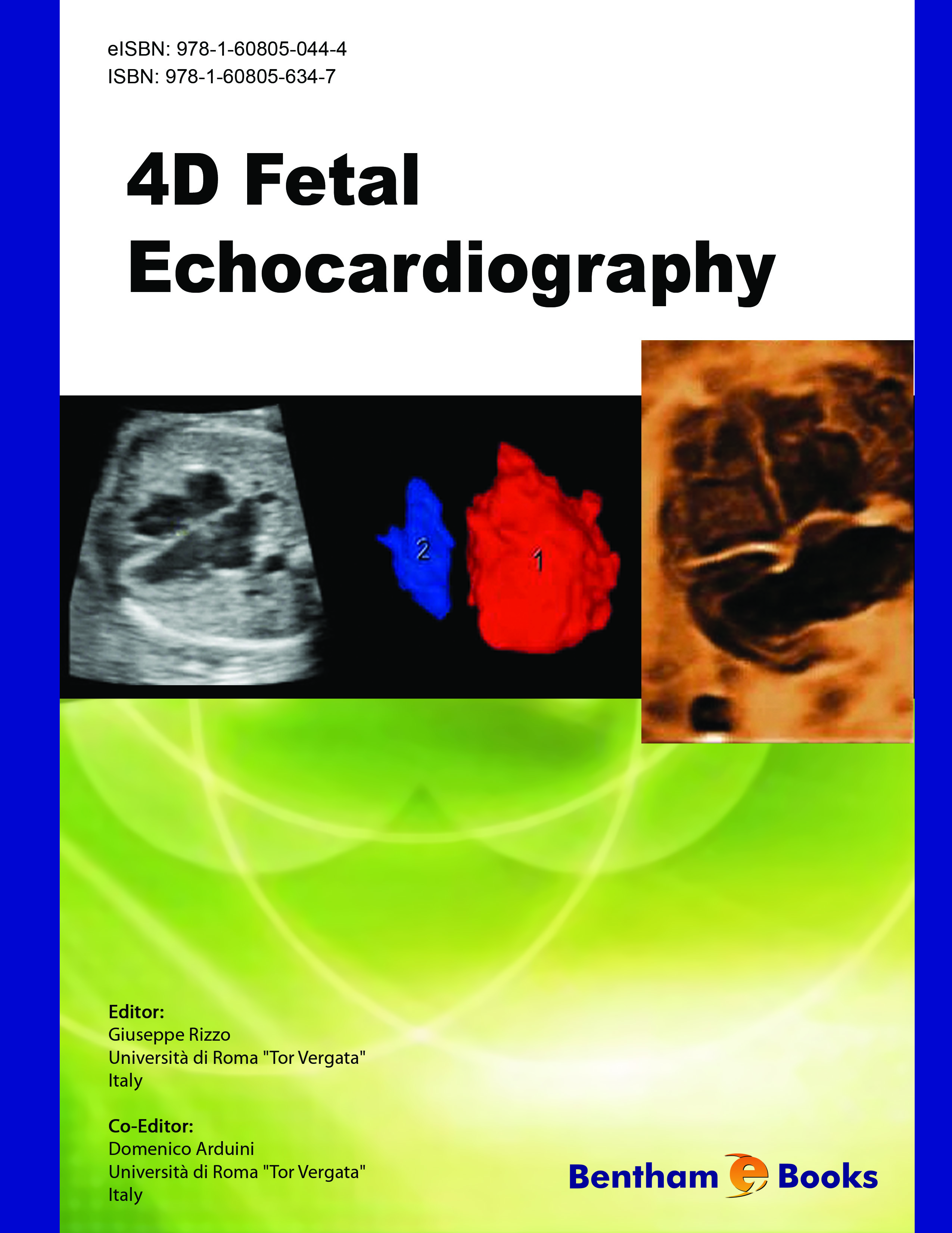
When Drs. Rizzo and Arduini asked me to write this preface, I was honored and thrilled to have the opportunity to introduce this outstanding work!
Examining the fetal heart has always presented a huge challenge for ultrasound specialists. When I started in ultrasound 30 years ago, no one knew how to perform or even attempt the examination of the heart. Fetal echocardiography has progressed very rapidly, fueled by advances in image quality as well as increasing focus of the ultrasound community on the need for a proper examination of the heart. Even today, the detection of congenital heart defects remains at the very bottom of the list of identifiable abnormalities on a standard fetal sonogram, and well under 50% of congenital heart defects are identified prenatally. Detection of congenital heart disease has lagged behind the successful detection of other abnormalities of the fetus despite our best attempts to teach the necessary skills. The fetal heart is a difficult organ to evaluate due toits very small size, complex anatomy, dynamic blood flow physiology, as well as rapid rhythmical movement.
The importance of evaluating the heart cannot be over emphasized. Heart defects are among the most common malformations in the developing fetus and many of these anomalies are part of complex syndromes such as aneuploidies. Heart anomalies that occur as isolated defects are among the most challenging to detect, but are particularly important due to the need to prepare for a potentially sick newborn at delivery. I remember back in the early 80's when I first started in fetal ultrasound, the heart remained an enigma for many years and it was only after I missed several congenital heart defects in a row that I took on the enormous task of understanding the fetal heart and how it works. Unfortunately back then we did not have any books or educational aids such as Drs Rizzo and Arduini's wonderful e-book to guide us in learning how to evaluate the fetal heart.
I have been impressed for many years with the careers of Dr. Rizzo and Dr. Arduini who have made enormous contributions in the field of fetal imaging and in particular fetal echocardiography. They have both published extensively on this subject and are extremely well qualified to write and edit such a book. Drs. Rizzo and Arduini have made significant contributions in fetal echocardiography over many years and have been instrumental in developing many of the Doppler aspects of the fetal heart evaluation. They have assembled an impressive group of authors here, and have edited a first class book on 3-D and 4-D of the fetal heart. They have understood that 3-D and 4-D volume imaging are necessary to evaluate the fetal heart due to its inherent complexity of motion, flow and anatomy.
The book is very well organized in multiple short chapters that are beautifully illustrated both with ultrasound images and diagrams. Initially, they introduce the subject with a chapter on the epidemiology of fetal heart defects and why they are important. Next are the basic chapters on screening and on normal anatomy, which are key for the beginners as well as for more seasoned examiner. The following chapters introduce 3-D and 4D ultrasound, starting with technical aspects on how to obtain a good image. Many practitioners are intimidated by the complexity of obtaining 3-D and 4-D images. The chapters that introduce the technical aspects of 3-D such as image display and manipulation of the image are all superb introductions on how to produce the best image. The book then focuses on 3-D and 4-D evaluation of blood flow using Doppler, a key subject in the evaluation of the heart. Additional chapters cover new technologies, such as the use of the matrix probe and the automated screening possibilities for the fetal heart. This volume includes the often forgotten venous anatomy of the heart and its connections, an extraordinarily valuable chapter. The chapters on anomalies are divided into septal defects, right heart, left heart, and cono truncal abnormalities and each chapter combines 2-D, 3-D/4-D and Doppler in the demonstration of these malformations. The book also describes the state of the art for 1st trimester echocardiography, followed by quantitative aspects of the function of the fetal heart including stroke volume and cardiac output. The last chapter involves other forms of imaging of the fetal heart, such as MRI.
This book, while very comprehensive, remains simple in its approach. It makes a very complex and rather intimidating organ accessible to both the novice and the seasoned examiner. Furthermore, this book is presented as an Ebook which is a very innovative format, awaited with great anticipation. Such novel media will challenge the primacy of bulky printed books. Many books will soon be available as Ebooks and carried as a small discrete thumb-drive or even down loaded perhaps on an electronic device such as a Kindle. Electronic versions of books are the future of our industry and Drs. Rizzo and Arduini have stepped up to the plate in choosing such a modern and practical display for their book.
In conclusion, I am very excited about this Ebook, not only because of its superbly organized, well illustrated and presented content, but also because as an Ebook it can be carried and propagated throughout our community much more easily than a hard copy book would be. The authors have succeeded admirably in their endeavor to produce what promises to be a outstanding resource on the examination of the fetal heart.
Beryl R. Benacerraf M.D.
Clinical Professor of OB GYN and Radiology
Harvard Medical School
Boston Massachusetts

