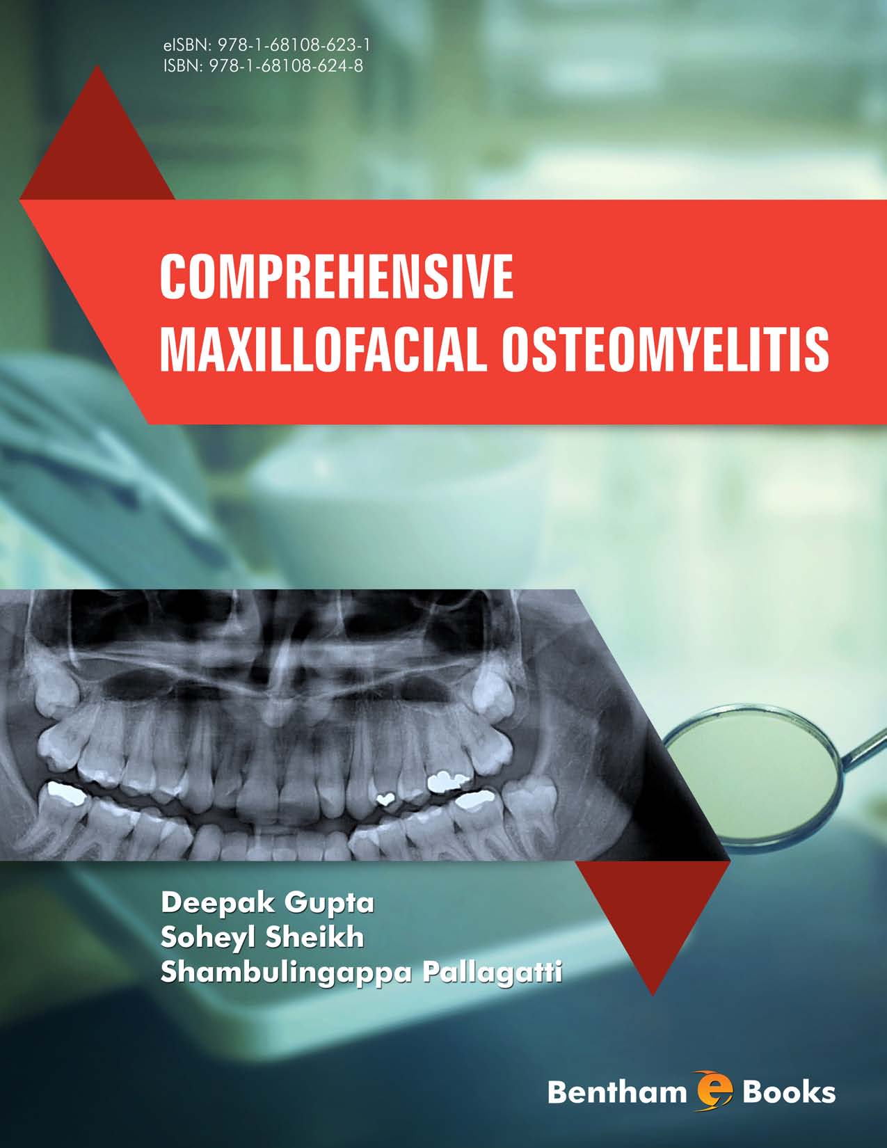
The word osteomyelitis is made up of a combination of two or three small words. In the Greek terminology, the word “OSTEON” means bone, the word “MYELOS” means marrow, and “ITIS” means inflammation. Henceforth, osteomyelitis may be considered as an inflammatory condition of the bone that usually begins as an infection of the medullary cavity, rapidly involves harversian systems and quickly extends to the periosteum area. It is considered to be a bacterial disease and was believed to be caused primarily by Staphylococcus aureus, Staphylococcus epidermidis, or hemolytic streptococci. But in recent years it has been found that 93% percent of the osteomyelitic infections of the jaws are polymicrobial with an average of 3.9 organisms per specimen. The most common predisposing factors for osteomyelitis to occur include irradiation, immunosuppressant treatment, malnutrition, and metabolic bone disease. It can also be precipitated by Paget’s disease of bone, fibrous dysplasia and bone tumors.
Osteomyelitis disease can involve any bone of the body. Bones of the jaws are also prone to develop this disease. Earlier before the advent of antibiotics, osteomyelitis of the jaws was frequently encountered as a result of odontogenic infections and was considered fatal. But with the advent of pertinent antibiotics, it was possible to control this disease. Literature reveals that the mandible is more commonly affected than the maxilla since the mandible is poorly vascularized as compared to the maxilla. The most common cause of osteomyelitis (OM) of the jaws is the spread of adjacent odontogenic infection. The second most common cause is the osteomyelitis occurring after a maxillofacial fracture, usually of the mandible.
The signs and symptoms of different types of osteomyelitis of jaws may include pain, which may be severe, throbbing and deep seated during initial stages. Later it may also lead to swelling, trismus, suppuration, fistula formation and fetid odour. It may further result in fibromyalgia, dysphagia, cervical lymphadenitis, paresthesia, fever, malaise and anorexia. Clinically pus exudation may be seen from around the neck of the teeth or from an extraction socket or from other sites. It starts as acute osteomyelitis. If the infection is not controlled, the process becomes chronic. This may even lead to loosening of teeth as well as sequestra formation. Untreated chronic osteomyelitis may even lead to pathological fracture.
Diagnostic imaging of osteomyelitis has been performed primarily through conventional radiographic modalities. These include intraoral periapical radiography, occlusal radiography and panoramic radiography. Advanced imaging modalities include computed tomography, magnetic resonance imaging (MRI) and bone scintigraphy. Since Maxillofacial osteomyelitis is chronic and debilitating disease, the main aim of the dental professionals is to diagnose the disease at early stages so as to initiate prompt treatment. Henceforth, there must be regular dental assessment and care.
ACKNOWLEDGEMENTS
For this Book my sincere thanks first of all goes to the Almighty, who brought me on this stage to meet my long cherished desire. In fact, it would have been impossible without His hidden unprecedented help and grace. I wish to express my sincere gratitude towards my guide, supervisor and teachers whom I would like to emulate.
I express my whole hearted and humble thanks to my beloved parents Mrs. Sunita Gupta & Mr. Ravinder Kumar Gupta whose unforgettable sacrifices and choicest blessings provided me the opportunity to be educated. My deep sense of obligation to them will ever remain for their constant inspiration and encouragement throughout the course of this task.
I am thankful to my wife Dr. Sonam Gupta, who showed her fervent interest, unusual generosity, accommodative attitude and gave me invaluable love, care and affection to accomplish this work.
I extend deep sense of gratitude to my brother Dr. Maqul Gupta and his wife Mrs. Mini Gupta for their unflinching moral support. They took personal interest and gave me constant encouragement since the inception of this work.
I owe gratitude to my mentors Dr. Amit Mittal (Vice-Principal, MMIMSR, Mullana) and Dr. Sanjeev Gupta (Professor and Head, Dept. of Dermatology, MMIMSR, Mullana) for their richness of vast experience and invaluable support throughout my academic carrier.
I would also like to thank my colleagues Dr. Amit Aggarwal, Dr. Ravinder Singh, Dr. Preeti Garg, Dr. Rahul Bansal, Dr. Johan Aps and Dr. Ibrahim Nasseh for their invaluable support and help throughout the process of completion of this task.
A special thanks to Dr. Debdutta Das (Principal, MMCDSR, Mullana) and Dr. Monika Das for their inspiring attitude and richness of vast experience.
CONFLICT OF INTEREST
There is no conflict of interest regarding publication of this book.
Deepak Gupta
Department of Oral Medicine and Radiology,
M.M. College of Dental Sciences and Research,
Mullana, Ambala, Haryana,
India
E-mail: drdeepak_26@rediffmail.com

