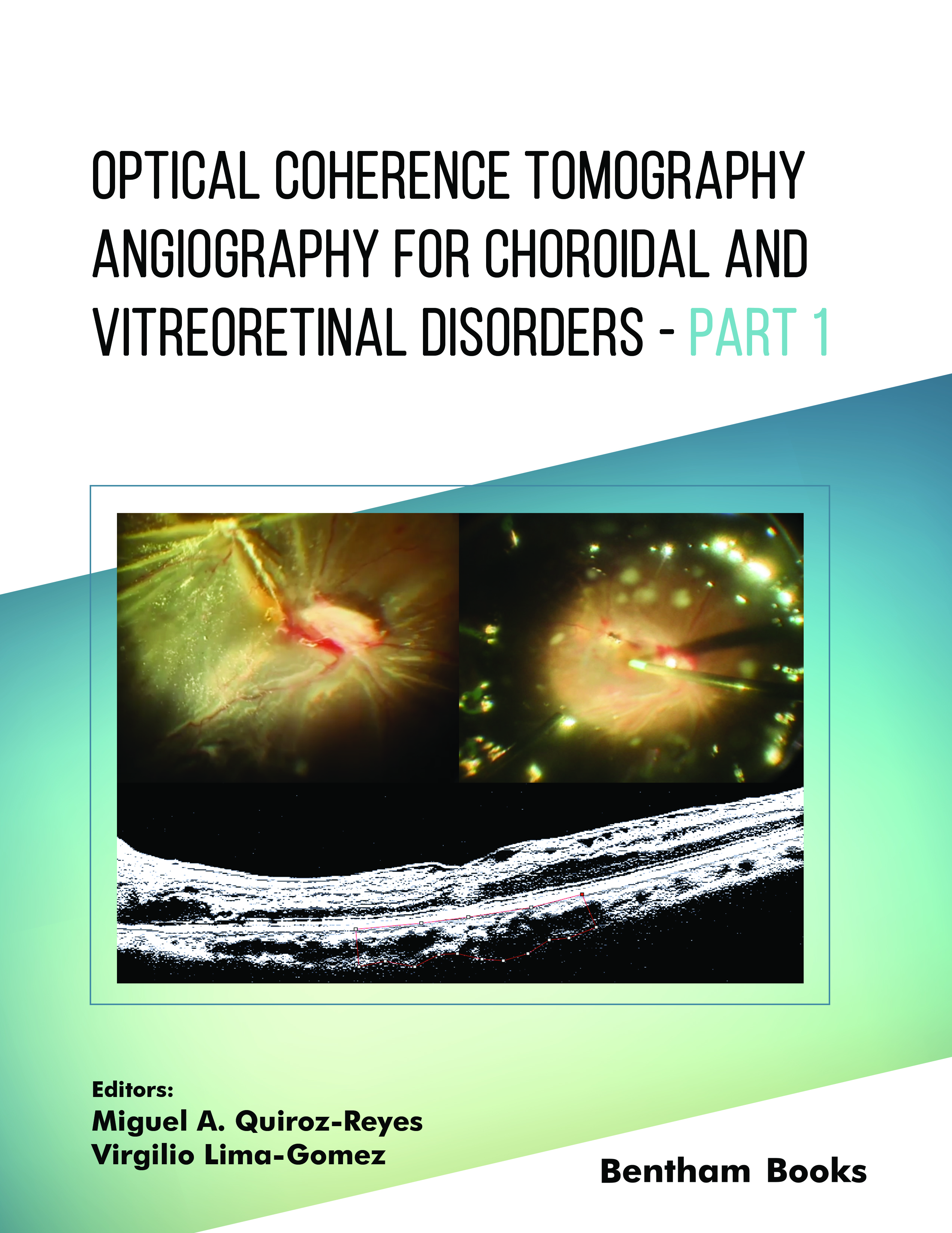
Optical coherence tomography angiography is one of the most important recent innovations in ophthalmology. The book you have in your hands represents the collaborative efforts of a select team of subject matter experts. This book aims to be a practical, patient-centered guide complemented with a clinical approach and demonstrative clinical cases to assist ophthalmologists and ophthalmology trainees in the evaluation of newly developed perfusion concepts and the diagnosis and management of patients presenting with a wide spectrum of diseases of the retina and choroid, as well as the role of perfusion parameters in the pathogenesis of diverse diseases. As mentioned briefly before, this book describes the journey from basic ophthalmology principles to the most sophisticated current aspects and advances that have resulted in the development of superb technological innovations. We have gone from fundus fluorescein angiography imaging to the evaluation of the perfusional indices of retinochoroidal structures using noninvasive and noncontact imaging techniques that allow a high histopathological correlation of structural tissue characterization with microvascular evaluation on tissue perfusion.
Written by leading international experts in the field, Optical Coherence Tomography Angiography for Choroidal and Vitreoretinal Disorders serves as a practical tool for daily work in a retina clinic, helping you through the first steps of perfusion investigation and clinical evaluation, correlation management and treatment decisions for these complex patients. Each chapter details distinct diseases of the retina or choroid, with a focus on signs and perfusion; optical coherent tomography is emphasized, and the chapters are illustrated with many multipaneled images, such that the book may be used as a reference for deciding on diagnostic and treatment options.
This book dissects the basics of angiography by optical coherence tomography and explains the differences in the clinical utility of optical coherence tomography as well as its complementarity. This gives us a broad explanation of the nomenclature and normal perfusional findings in healthy populations.
Several chapters explain macular perfusional findings in different vitreoretinal and choroidal pathologies, including vascular entities commonly seen in daily practice, such as diabetic retinopathy, hemorrhagic and ischemic infarctions of the retina due to vascular disorders, and choroidal pathological neovascularization; most importantly, perfusion parameters are evaluated by quantification and binarization of the different vascular plexuses at the retinal and choroidal level. Additionally, certain tractional entities are evaluated from the point of view of their microstructural findings and perfusional postoperative outcomes, associating them with the final vision.
Some chapters deal with new antivascular endothelial growth factor molecules and new extended-release delivery devices and provide a comparative evaluation of the therapeutic effect on perfusion. In this way, multiple complex pathological disorders of the retina and choroid are more efficiently diagnosed, followed by natural and treated medical or surgical evolution according to the specific cause and consequently, as mentioned before, monitored in response to specific treatments.
We hope that this book, from a multitude of experts, contributes pertinently to academia and achieves the objective of serving as a guide both in the diagnosis and clinical decision-making that those of us who are dedicated to the difficult but beautiful and challenging practice of clinical and surgical retina care perform on a daily basis.
Miguel A. Quiroz-Reyes, MD
Oftalmologia Integral ABC, Retina Department
Medical and Surgical Assistance Institution (Nonprofit Organization)
Affiliated with the Postgraduate Studies Division
National Autonomous University of Mexico
Mexico City, Mexico
&
Virgilio Lima-Gomez, MD
Ophthalmology Service, Hospital Juarez de Mexico
Public Assistance Institution (Nonprofit Organization)
Mexico City, Mexico

