Editor: Cleverson Teixeira Soares

Histopathological Diagnosis of Leprosy

eBook: US $59 Special Offer (PDF + Printed Copy): US $132
Printed Copy: US $103
Library License: US $236
ISBN: 978-1-68108-800-6 (Print)
ISBN: 978-1-68108-799-3 (Online)
Year of Publication: 2021
DOI: 10.2174/97816810879931210101

Introduction

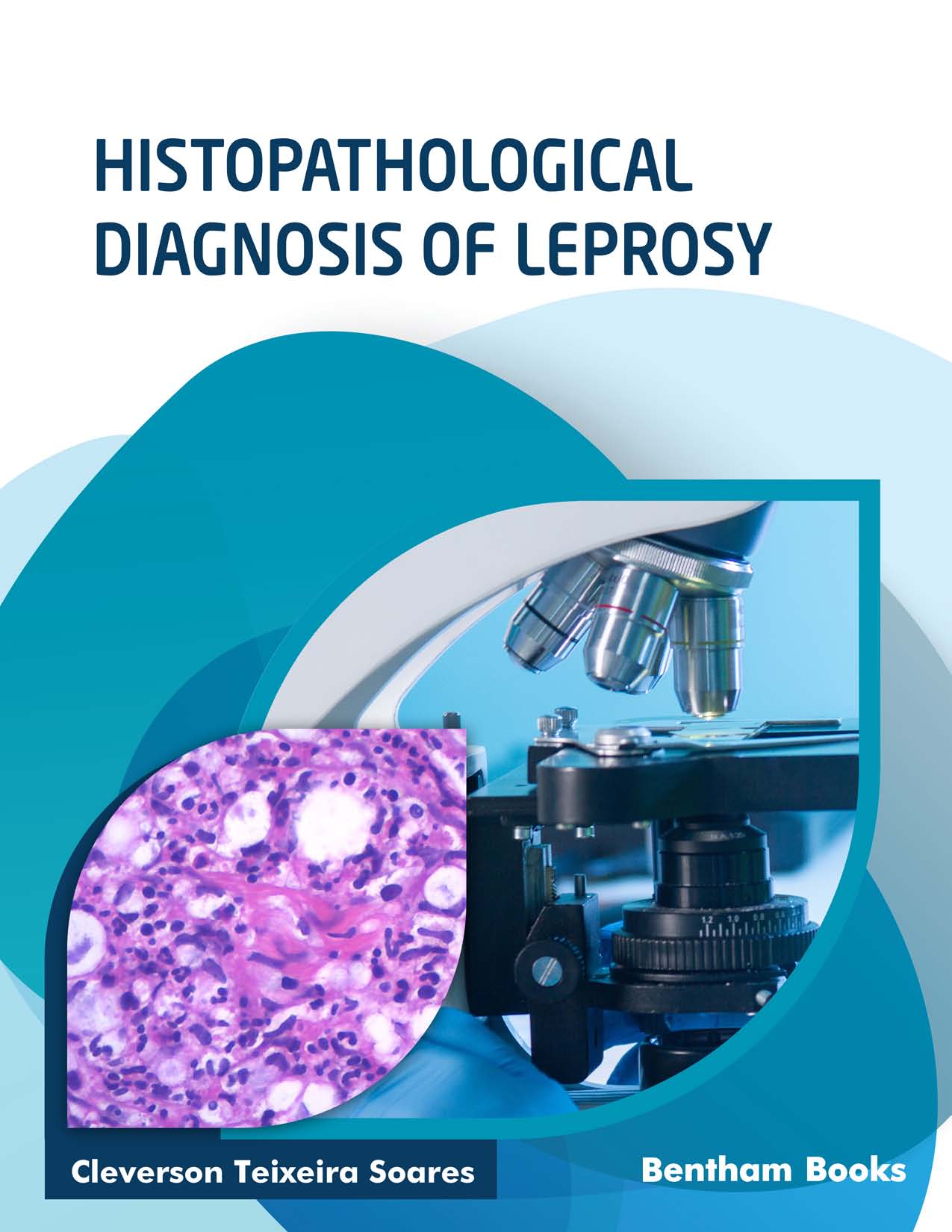
Histopathological Diagnosis of Leprosy, is a comprehensive guide to the medical pathology of Hansen’s disease, which is a complex and clinically challenging infection caused by Mycobacterium leprae. Readers will find 8 chapters on key topics on the subject including general aspects of leprosy, different forms of leprosy (polar, borderline, etc.), reaction types and complications. The information presented in the handbook will equip the reader with the knowledge required to identify the disease in patients and perform differential diagnosis where required.

Key Features:

-8 chapters dedicated to key topics about leprosy and its diagnosis.

-More than 200 figures featuring over 1000 clinical and histopathological photographs

-Complete information about differential diagnosis and reaction phenomena

-includes a section dedicated to special and complicated cases

-References for further reading

-Brings the expertise of renowned physicians to the reader

The detailed presentation of the book is of great value to both healthcare professionals (pathologists, dermatologists, physicians) who are involved in the care of leprosy patients, and medical residents who are seeking information about the disease as part of their medical training.

Preface

Leprosy remains one of the biggest public health problems in many countries. It is a complex disease in several aspects, including clinical, histopathological, and molecular. This book is written to help different professionals (medical students, physicians, pathologists, researchers, and others) who handle this disease daily or are interested in it to understand the disease from the perspective of pathology. The histopathological characteristics of leprosy throughout its spectrum, reaction phenomena, and clinical presentation types are crucial for understanding the pathophysiology of the disease and guiding clinical conduct and research accordingly.

The book is organized into eight sequential chapters that allow the readers to gradually immerse themself in different histopathological aspects of leprosy. The clinical features discussed are accompanied by corresponding histopathological images presented as photo panels, along with key points that assist in understanding the described characteristics. Common histological sections stained by hematoxylin-eosin are complemented with those stained by Fite–Faraco to identify the different types of cells or tissues parasitized by Mycobacterium leprae . Immunohistochemical findings are used to clearly illustrate some characteristics observed in common histological sections. In some cases, more than one staining technique was used for the same histological section (for example, immunohistochemistry and Fite–Faraco), thereby contributing to a better understanding of the histopathological characteristics of the disease.

The interpretation of the histopathological characteristics of leprosy lesions is somewhat challenging. Each diagnosis depends on the close correlation between the findings of histopathology, bacilloscopy examination, and clinical data. In this context, histopathology is of great importance to confirm or dismiss a certain hypothesis and can guide teams to accurately interpret data and choose the best clinical and therapeutic conduct.

This book is intended to be a supplement to the leprosy literature found in journals and textbooks, offering the readers a background on leprosy-related histopathology that can improve their understanding of this disease.

Finally, I express deep gratitude toward the different support teams of the Lauro de Souza Lima Institute, especially that of the Laboratory of Anatomic Pathology, which contributed decisively to the conclusion of this book and to Dr. Cássio C. Ghidella for the numerous clinical photos that illustrate all chapters.


Cleverson Teixeira Soares
Laboratory of Anatomic Pathology
Instituto Lauro de Souza Lima (ILSL) and
Institute of Pathology of Bauru (ANATOMED) Bauru
Brazil

RELATED BOOKS

.COVID 19 – Monitoring with IoT Devices.
.Recent Advances in the Application of Marine Natural Products as Antimicrobial Agents.
.Recent Trends and The Future of Antimicrobial Agents - Part 2.
.COVID-19: Origin, Impact and Management (Part 1).