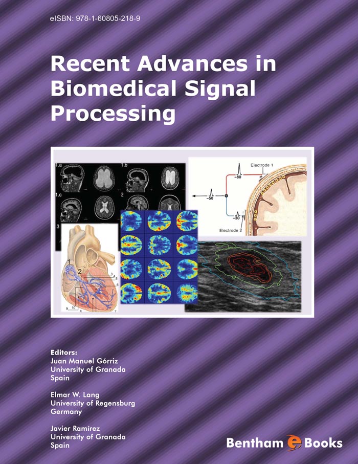
Few years ago, processing of biomedical signals was mainly concentrating on filtering of signals for removing noise and power lines interference; spectral analysis to understand the frequency characteristics of signals; and modeling for feature representation and parameterization. Biomedical signal processing is a rapidly expanding field with a wide range of applications, from the construction of artificial limbs and aids for disabilities to the development of sophisticated medical imaging systems. These include ultrasound scanners, magnetic resonance imaging scanners and positron emission tomography (PET and SPECT). X-ray systems have also improved and are widely used for many purposes such as mammograms.
On the other hand, acquisition and processing of biomedical signals has become more and more important to the physician. The main reasons for this development are the growing complexity of the biomedical examinations, the increasing necessity of comprehensive documentation and the need for automation in order to reduce costs. Recent trends have been toward quantitative or objective analysis of physiological systems via signal analysis. Analysis of signals accomplished by humans has many limitations, therefore, computer analysis of these signals could provide objective strength to diagnoses; however, the development of an algorithm for biomedical signal analysis is a significant challenge. Different techniques can be used to analyze a biomedical signal, these include: filtering, adaptive noise cancellation, and pattern recognition (to differentiate between abnormal and normal physiological signals). Medical image processing, using techniques such as X-ray and MRI, can be viewed as multi-dimensional signal processing.
In addition, medical practitioners are increasingly using computer-based medical systems to collect, store, and process digitized biological signals. The signals they collect and process via these systems include bioelectric potentials, such as those generated by the heart (ECG or electrocardiogram), brain (EEG or electroencephalogram), or skeletal muscles (EMG or electro-myogram). Such signals include nonelectric signals that might be transduced and then recorded (for example, breath sounds or speech waveforms), and biomedical images from ultrasound, Xray, AT scan, MRI, SPECT, PET, etc. These signals are often interpreted heuristically by medical practitioners, and the need for sophisticated algorithms for processing, coding, and automatically interpreting the information these signals contain is increasing. Among the advantages of automated processing are objectivity, reliability, repeatability, and speed. Of course, as biomedical signal processing algorithms gain sophistication, more computational resources are needed, while most biomedical signal processing must be done in or near real time. The volume of data is often high, computational resources are often scarce, and the cost of resources and computing time is important. Thus, it is necessary to simultaneously consider not only whether a particular technique is useful but also how it might be efficiently mapped to special-purpose hardware to be included in a computer-based medical system. The transition from uni-processor to multiprocessor architectures is mandatory to achieve real-time performance in computerbased medical systems for biomedical signal and image processing.
This e-BOOK will cover biomedical signal processing as used in both therapeutic and diagnostic instrumentation. A number of current research projects will also be outlined with emphasis on intelligent medical image diagnosis. We would like to express our gratitude to all the contributing authors that have made a reality this book. We would like to also thank Dr. Principe for writing the foreword and Bentham Science Publishers, particularly Manager Sara Moqeet, for their support and efforts.
Juan M. Górriz, Javier Ramírez and Elmar W. Lang
University of Granada, Spain University of Regensburg, Germany

